+5411 4493 7422 | [email protected]

Diagnosis

Avatar
pablo
7 years ago

Definition

Mucocutaneous lymph node syndrome (MLNS), or Kawasaki disease, is a multisystemic vasculitis of unknown etiology and immunological pathogenesis, that manifests in babies and young children; 85% of affected children are under the age of 5. It has a well defined clinical criteria for diagnosis and its course is self-limited to 60 days.

Cardiac compromise is present in 40% of the cases, and 20% of the patients show coronary alterations. Mortality rate ranges from 0.2 to 1% due to myocarditis or acute myocardial infarction (AMI), secondary to similar coronary arteritis or identical to childhood polyarteritis nodosa’s pattern.

Historical Overview

The disease is named after Dr. Tomisaku Kawasaki who first described the case in Japan, in 1967. He called it mucocutaneous lymph node syndrome and described the clinical criteria for diagnosis that remain valid to this day. It was not until 1974 that the first series of 50 cases would appear in american literature, and two years later, Drs Melish and Hicks would publish the first cases in the United States.

In Argentina, the first documented case appeared in the year 1976 and it corresponded to a patient of the Children’s Hospital «Ricardo Gutiérrez», where it was presented in the scientific conferences, 10 years after its first description. In 1979, Kato published a paper in Pediatrics about the treatment with acetylsalicylic acid and the recommendation to avoid corticoids.

In 1984, Furusho’s publication in Lancet would appear, and two years later, Newburger’s in the New England Journal of Medicine, where they would share their experiences with high doses of intravenous gamma globulin, thus establishing the bases for the current treatment and reducing the impact in coronary affectations.

However, this disease already existed, since in 1870, St. Bartholome’s Hospital in London examined the heart of a deceased child diagnosed with «Scarlet Hydropsy» who had three coronary aneurysms as well as characteristic pathological changes.

In June 2020, Dr. Tomisaku Kawasaki passed away, leaving the legacy of his wisdom.

Main criteria for diagnosis

Ícono Temperatura

A fever that lasts more than 5 days and doesn’t respond to antipyretics or antibiotics.

Ícono Ojo

Bilateral conjunctival injection without suppuration.

Ícono Pies

Changes in the extremities, swollen and/or colored hands and feet:
– Acute phase (0-12 days): erythema of the palms and soles accompanied by brawny edema.
– Subacute phase (12-45 days): desquamation of the tips of fingers and toes.
– Convalescent phase (45-60 days): Beau’s lines (deep transverse grooves across the nails). Helpful for a retrospective diagnosis.

Ícono Manos

Skin changes, red outbreaks without vesicles or blisters. Irritability, restlessness, abdominal pain, diarrhea.

Ícono Boca

Mouth changes. Red, cracked lips, red tongue, no ulcers: erythema, edema and/or cracked lips, strawberry tongue, cheilitis and diffuse congestion of the pharynx and oral cavity.

Ícono Cuello

Unilateral or bilateral non-purulent acute cervical adenopathy.

The diagnosis for KD requires the presence of 5 items, and ruling out other pathologies as well.

In the presence of coronary aneurysms, the diagnosis can be done with 3 or 4 items.

Incomplete and/or atypical cases appear in children of all ages, but more frequently in children under 2, and older than 10 years old. They require a clinical, electrocardiographic and echocardiographic follow-up as «possible cases».

Kawasaki shock syndrome (KSDS) in 2 to 5% of cases in children or older than 6 years and adolescents. Indistinguishable or similar to Covid 19-associated multisystemic inflammatory syndrome.

Images

Suspicious cases follow-up algorithm

Kawasaki Disease Diagnosis
References
Taken from: Vainstein E., Baleani S. Enfermedad de Kawasaki casos incompletos o atípicos. Estrategia diagnóstica. Revista del Hospital de Niños (B. Aires) 2007; 49(222): 93-98.
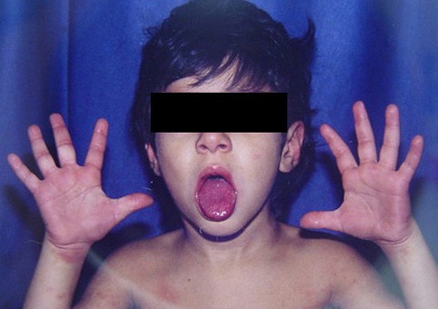
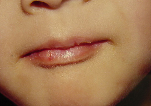
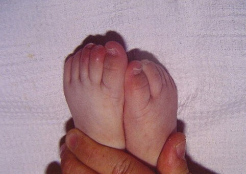
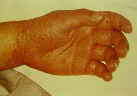
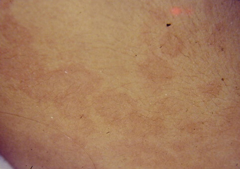
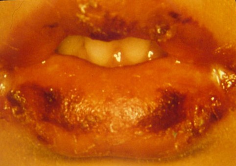
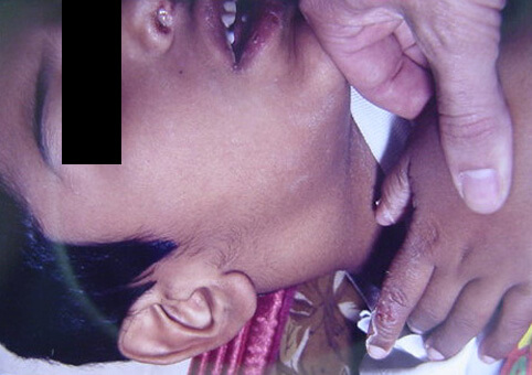
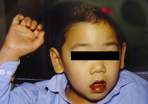
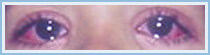
1 Children ≤6 months old, after ≥7 days of unexplained fever: must undergo lab tests and if there is evidence of systemic inflammation an echocardiogram must be done, even in the absence of clinical criteria. 2 See Diagnosis criteria. Characteristics that suggest another disease: exudative conjunctivitis, exudative pharyngitis, mild intraoral lesions, bullous exanthems or generalized adenopathy. Consider alternative diagnoses. 3 Laboratory criteria: albumin ≤3 g/dl, anemic for such an early age, increased alanine aminotransferase, platelet recount ≥450.000/mm3 after 7 days, white blood cells recount ≥15.000/mm3 and urine with ≥10 white blood cell/field. 4 Can be treated before the echocardiogram. 5 Echocardiogram: consider as positive the presence of perivascular refractivity, reduced left ventricular function, mitral regurgitation, pericardial effusion, lumen diameter of coronary arteries >3 mm in <5-year-olds, and >4 mm in >5-year-olds; if the diameter of a segment measures ≥1,5 times the adjacent segment or in relation to the aorta, or if there are irregularities in the coronary lumen. The echocardiogram must be done by a trained professional. 6 Pathological ECG: arrhythmia, tachycardia, prolonged PR interval, low QRS voltage, changes in ST-T waves, abnormal Q waves, repolarization disorders. 7 If the echocardiogram or the ECG are positive, treatment must be administered within 10 days since the fever started, and after 10 days, patients with lab (C reactive protein, erythrocyte sedimentation) and clinical signs that indicate inflammation. 8 Typical desquamation starts under the nail bed of fingers and soon after, toes. CRP: C reactive protein. ESR: erythrocyte sedimentation rate. Echo: echocardiogram. KD: Kawasaki disease.

Associated clinical criteria

Sterile pyuria, urethritis, myo-peri-pancarditis, arthritis, arthralgia, irritability, aseptic meningitis, abdominal pain, compromised liver, gallbladder hydrops, diarrhea, obstructive jaundice.

Clinical history/background

Age, sex. Stage of the disease. Onset and disappearance of symptoms and signs. Received medication (it is important to establish if the patient received corticoids since they increase coronary compromise). Exposure to infecto-contagious and/or toxic diseases.

Hospitalization criteria

All children diagnosed with Kawasaki disease that haven’t received treatment.

Admission will not be considered for patients in a convalescent stage, that have good health and no coronary or cardiac compromise, and can comply with a strict out-patient follow-up.

Admission testing

Physical exam: full clinical examination, with special focus on skin, circulatory and osteoarticular systems. Examine scalp, armpits and groin, and areas where aneurysms tend to appear (feel all arterial pulses).

Additional exams: Hemogram with platelet count, erythrocyte sedimentation rate (ESD), C-reactive protein (CRP) weekly or biweekly until normalization. Hepatograms (BiT and Direct, GOT, GPT, gamma glutamyl transpeptidase), Proteinogram, Ionogram, Troponin I, ProBNP, Complete urine, Hemocultures, Serologies. Chest teleradiography. Electrocardiogram. Two-dimensional transthoracic cardiac ultrasound, in order to evaluate cardiac and/or coronary involvement. It should be repeated at 14-28-60 days in search of cardiac and / or coronary alterations. Abdominal ultrasound (vesicular hydrops). Ophthalmologic examination with slit lamp (keratitis, uveitis).

Treatment

The treatment points to four main purposes:

To quickly reduce the inflammatory process.

To regulate the immune system.

To prevent cardiovascular lesions.

To prevent thrombosis.

Supportive care

Rest, hydration.

Specific

Period: 0 to 12 days.

Aspirin: 50-100 mg/kg/day, oral. 4 doses to reduce inflammatory signs.

Intravenous gamma globulin of 2 g/Kg/dose. Continue slowly from a 12 hour constant infusion a day to prevent aneurysms. The infusion speed during the first two hours must be half of the amount calculated for an hour to evaluate possible side effects like: rash, arterial hypertension or cardiovascular compromise. In case of side effects, antihistamines can be administered or the infusion rate can be extended to 18-24 hours accordingly.

Retreatment: a second series of gamma globulin at 1-2 grams/kg/dose is considered if clinical symptoms reappear or fever persists for more than 48 hours after the first dose, with elevated CRP or ProBNP, indicating active disease or persistence of inflammatory activity.

Corticoids use: the use of intravenous methylprednisolone pulses at 30mg/Kg/dose in 2 hours for three consecutive days is proposed in the case of disease refractory to treatment and retreatment with gamma globulin with persistence of clinical symptoms and altered laboratory. After consultation with the specialized service.

Monoclonal antibodies (moAb) use: Infliximab, Anakinra, Tocilizumab, in patients who do not respond to previous therapies. Consult the specialized service.

Peripheral gangrene: when distal perfusion disorders of hands and feet (Raynaud’s) are evidenced, the use of Prostacyclins, Anticoagulants (Low Molecular Weight Heparin), Methylprednisolone pulses and/or monoclonal antibodies should be considered. Consult a specialized service.

SSKD: Consultation with specialist, admission to ICU. Rapid and sequential therapy with gamma globulin and corticosteroids or monoclonal antibodies.

Period: 12 to 60 days.

Without cardiac compromise:

ASA 3-5 mg/Kg/day, for 2 months in one morning dose (antiaggregant effect).

With cardiac compromise:

ASA 3-5 mg/Kg/day. Depending on the patient’s evolution it will be continued indefinitely.

Dipyridamole (Persantine tablet) 3-5 mg/Kg/day in 2-3 doses combined with ASA, in patients with severe coronary disease or severe thrombocythemia (platelet counts over 700.000).

Surgical: according to the cardiovascular surgeon, by-pass, Stent, valve replacement.

Period: after 2 months.

Patients without cardiac compromise:

Ambulatory clinical control. Annual cardiac checkup.

Patients with cardiac compromise:

Medical treatment: ASA combined or not with dipyridamole. Monitoring of coronary risk’s factors (total cholesterol, HDL, obesity, tobacco screening, arterial hypertension, sodium intake control, etc.).

Treatment and cardiac follow-up: taking into account the after-effects: cardiomyopathy, giant aneurysms, coronary insufficiency. Clinical-echographic follow-up. Each case will deem coronary perfusion and ergometrics tests accordingly. Specialists along the cardiology department will evaluate each case regarding the need for and possibility of a coronarography.

For more information, see the bibliography.